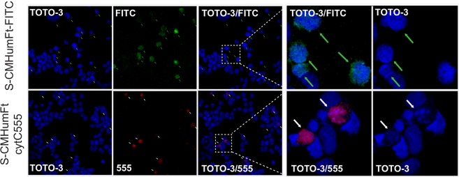
Figure 3.
Confocal microscopy of APL NB4 cells treated with S-CMHumFt-FITC and S-CMHumFt-Cyt C 555. Cytochrome C labeled with AlexaFluor 555 is delivered into the APL NB4 cell line by S-CMHumFt leading to apoptotic cell death. NB4 cells were cultured in the presence of 150 μg/mL S-CMHumFt-FITC (upper panels) or of 150 μg/mL S-CMHumFt loaded with cytochrome C-AlexaFluor 555 (lower panels) for 24 hours and analyzed by confocal microscopy, after labeling the DNA with the TOTO-3 dye. All the cells that internalized detectable amounts of Cyt C 555 presented apoptotic nuclei as indicated by nuclear condensation (white arrows). On the contrary, even the cells that internalized high amounts of S-CMHumFt-FITC alone showed vital, uncondensed nuclei (green arrows). The panels on the right show an enlargement of the cells into the insets.

