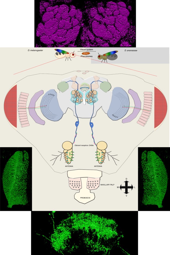
Fig. 5.
Model for the neural circuitry of dialect training in Drosophila. Inter-species communication and dialect learning is dependent on the presence of multiple cues between D. melanogaster and D. ananassae. These cues are, in part, olfactory, visual, and ionotropic in nature. In D. melanogaster, we have identified multiple brain regions and neurons that contribute to the dialect training. Regions we identified to be involved in dialect training—the optic lobe (blue), mushroom body (orange), antennal lobe (teal), lateral horn (green), fan-shaped body (green/white), and ellipsoid body (light green). For the antennal lobe, we identify the D glomerulus, innervated by Or69a olfactory receptor neuron (ORN). For the fan-shaped body, we identify region 5 as necessary for dialect training. For the optic lobe we identify the motion-detecting circuit as necessary by isolation of the L4 neurons. Flanking the model, super-resolution microscopy with a 3D render is provided of the key regions of the fly brain that were investigated—region 5 of the fan-shaped body, the L4 neurons, and the antennal lobe. Super-resolution images without 3D render are located in Supplementary Fig. 20

