Fig. 1.
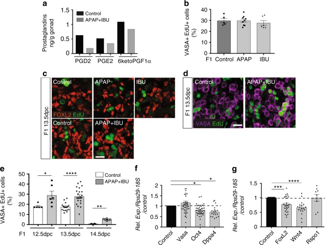
In utero exposure to APAP and NSAIDs increases female germ cell proliferation. a Dosage of PGD2, PGE2 and 6-keto-PGF1α (PGI2) by mass spectrometry in F1 13.5 dpc control and APAP + IBU-exposed ovaries (n = 1 pool of 200 and 220 gonads from 15 control and 15 treated pregnant females, respectively). b Quantification of VASA+ EdU+ cells in F1 control and APAP or IBU-exposed ovaries; data are the percentage of proliferating EdU+ cells among all VASA+ cells (n = 20–25 gonads from n = 12 litters). c, d Representative immunofluorescence microscopy images of F1 13.5 dpc ovaries from embryos in utero exposed to ethanol (Control), APAP, IBU or APAP + IBU between 10.5 and 13.5 dpc and pulsed with EdU at 13.5 dpc; FOXL2 (granulosa cell marker) in red (c), VASA (germ cell marker) (d) in purple and EdU in green (scale bars, 20 μm). e Percentage of proliferating EdU+ cells among all VASA+ cells in F1 control and exposed ovaries at 12.5, 13.5 and 14.5 dpc (EdU pulse at 12.5, 13.5 and 14.5 dpc, respectively) (n = 16–20 gonads from n = 8 litters). f, g Expression analysis of Vasa, Oct4, Dppa4 (f), Foxl2, Wnt4 and Rspo1 (g) in F1 exposed ovaries, normalised to Rps29 and 18S expression and presented as percentage of the expression in control 13.5 dpc ovaries (set to 1). Values are the means ± SEMs; *P < 0.05, **P < 0.01, ***P < 0.005, ****P < 0.001 (e–g)

