Fig. 5.
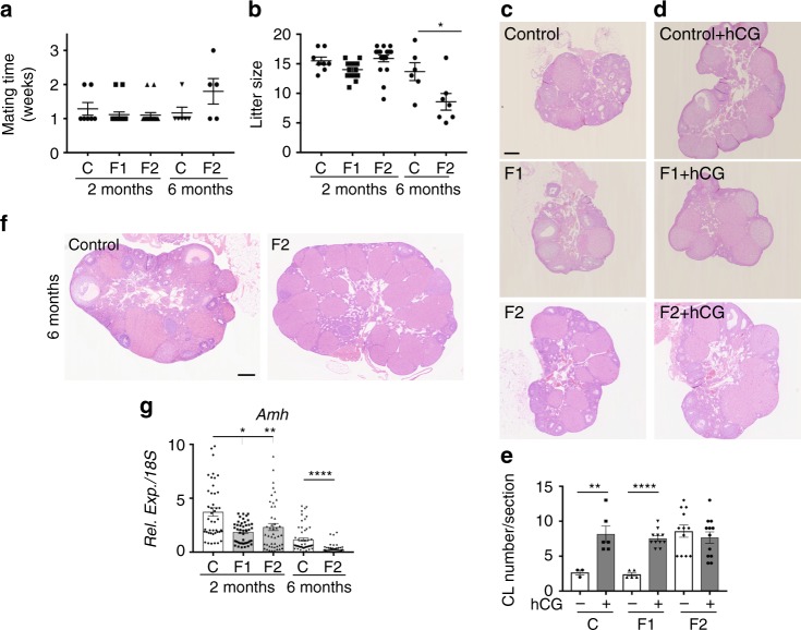
F2 females show accelerated ovarian aging and are subfertile. a, b Fertility of 2-month- and 6-month-old females was determined in terms of time to mating (a) and litter size (b) (n = 6–19 for each data point) *P < 0.05. c, d, f Histological analysis of 2-month-old ovaries stimulated (d) or not (c) by hCG and of 6-month-old ovaries (f). All sections were stained with haematoxylin and eosin. Scale bars = 200 μm. e Corpus luteum (CL) number measured in three representative sections of each ovary (n = 10 ovaries). **P < 0.01, ***P < 0.005 (hCG treated vs controls). g Amh expression levels in 2-month- and 6-month-old F1 and F2 ovaries, compared with their respective controls. *P < 0.05, **P < 0.01, ****P < 0.001

