Figure 1.
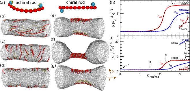
Membrane tube deformation induced by chiral and achiral protein rods without direct attraction between the rods at Rcyl/rrod = 1.31 and N = 4800. (a) Protein models for achiral and chiral crescent-shaped rods. (b–g) Snapshots at (b) Crodrrod = 0, (c) 1.5, and (d) 2.3, and (e,f) 3.3 for the chiral rods; and (g) Crodrrod = 3.3 for the achiral rods. The membrane particles are displayed as transparent gray spheres. (h,i) Fourier amplitudes of (h) rod densities and (i) membrane shapes. The solid and dashed lines represent the data for REMD of the chiral and achiral protein rods, respectively. The circles and squares with solid lines represent the qθ and qz modes, respectively, for the canonical simulations of the helical rod-assembly as shown in (f). The Fourier amplitudes are normalized by the values at Crod = 0 (denoted by the superscript *). The error bars are displayed only for the canonical simulations. The errors in REMD are smaller than the thickness of the lines.

