Fig. 2.
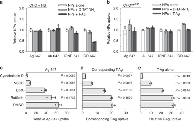
Mechanistic studies of bystander uptake. a Bystander uptake is completely abolished by HS. CHO cells were incubated with HS prior to the addition of the indicated bystander NPs (x-axis) alone, or with D-TAT-NH2 or T-Ag. The fluorescence intensity of bystander NPs per cell was quantified by flow cytometry and normalized to that of bystander NPs alone (y-axis). Data presented here are mean ± s.d. of three independent experiments (n = 3). b The indicated bystander NPs (x-axis) were incubated with CHOpgs745 cells with D-TAT-NH2 or T-Ag, and bystander NPs uptake (y-axis) was quantified as described in a. Data shown are mean ± s.d. of three independent experiments (n = 3). c–e CHO cells were pre-treated with indicated MP inhibitors dissolved in DMSO (y-axis) before incubating with T-Ag and Ag-647. The fluorescence intensity of AgNPs was quantified by flow cytometry and normalized to that of Ag-647 (for Ag-647 bystander uptake) or Ag-555 (for T-Ag uptake) alone (x-axis). c bystander uptake of Ag-647; d corresponding T-Ag uptake in c; e the uptake of T-Ag alone. All quantified data were analyzed using one-way ANOVA with Tukey’s multiple comparisons test comparing DMSO versus other MP inhibitors and are expressed as mean ± s.d. of three independent experiments (n = 3). One-way ANOVA, c F = 91.712, P = 0.0085; d F = 829.55, P = 0.0001; e F = 641.67, P < 0.0001. Source data are provided as a Source Data file

