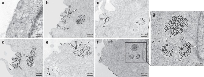
Fig. 3.
TEM analysis of endocytic structures for bystander uptake. CHO cells were incubated with Au50 alone (a) or Au50 + T-Au (b–g) in DMEM medium for 1 h before being fixed and processed for TEM imaging. The triangle arrows highlight the Au50 particles in the corresponding images. The Au50 particles were found together with T-Au in macropinosome-like endocytic vacuoles (>200-nm in diameter) (b–d), early endosome (e, bottom left), and late endosomes and lysosomes (e, g). Scale bars, 200 nm (except in f, in which is 500 nm). Three independent experiments (n = 3) were performed and representative TEM images are shown here. Source data are provided as a Source Data file

