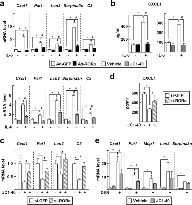
Figure 2.
RORα regulates gene expression of APPs. (a,b) Primary mouse hepatocytes were infected by Ad-GFP or Ad-RORα for 24 h and then treated with 20 ng/ml IL-6 for 24 h (top). Or primary mouse hepatocytes were treated with 50 μM JC1-40 for 24 h in the presence or absence of 20 ng/ml IL-6 for 24 h (bottom). The mRNA levels of APR markers such as Cxcl1, Pai1, Lcn2, Serpina3n, and C3 were analyzed by qRT-PCR (a). Culture media were collected and the level of CXCL1 was detected by ELISA (b). (c,d) Primary mouse hepatocytes were transfected with si-GFP or si-RORα for 24 h and then treated with 50 μM JC1-40 for additional 24 h in the presence of 20 ng/ml IL-6. The mRNA levels of APR markers such as Cxcl1, Pai1, Lcn2, Serpina3n, and C3 were analyzed by qRT-PCR (c). Culture media were collected and the level of CXCL1 was detected by ELISA (d). (e) JC1-40, 20 mg/kg BW/day, was administered orally for 3 days to the mice and then the mice were i.p. injected with 100 mg/kg BW DEN for 2 days before sacrificed. The hepatic mRNA levels of APR markers were analyzed by qRT-PCR. *P < 0.05; #P < 0.05 (n = 3) for (a–d) and (n = 3–5) for (e). The data represent mean ± SD. Representatives of at least three independent experiments with similar results are shown.

