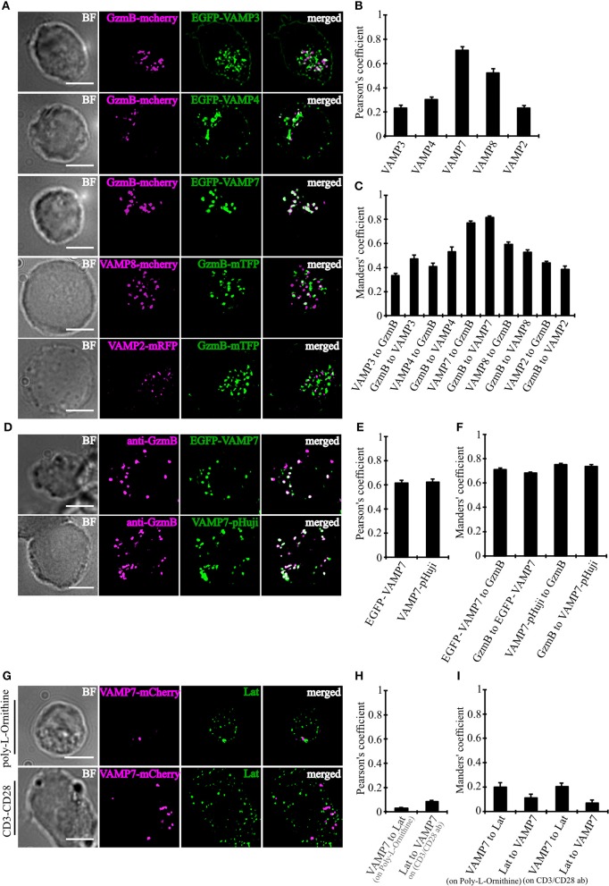
Figure 2.
VAMP7 shows a high degree of co-localization with cytotoxic granules. (A) SIM images of bead-stimulated human CD8+ T cells co-transfected with either granzyme B-mCherry or granzyme B-mTFP along with the v-SNAREs mVAMP2, VAMP3, VAMP4, VAMP7, and VAMP8 constructs. Overlay of both channels displayed that only VAMP7 co-localizes with the lytic granule marker granzyme B. (B) Pearson's and (C) Manders' overlap coefficients for co-localization of VAMP3 (n = 16), VAMP4 (n = 14), VAMP7 (n = 13), VAMP8 (n = 9), and VAMP2 (n = 14) with granzyme B are given in the text. (D) Bead-stimulated human CD8+ T cells transfected with EGFP-VAMP7 or VAMP7-pHuji and immunolabeled with Alexa647-conjugated anti-granzyme B antibody. (E) The corresponding Pearson's and (F) Manders' overlap coefficients for colocalization of EGFP-VAMP7 (n = 12) or VAMP7-pHuji (n = 11) with granzyme B are given in the text. Data are shown as mean ± SEM. Scale bar, 5 μm. (G) Anti CD3/CD28 bead-stimulated human CD8+ T cells transfected with VAMP7-mCherry and incubated on either poly-L-ornithine or anti CD3 antibody coated coverslips for 15 min, fixed and immunolabeled with LAT antibody. (H) The corresponding Pearson's and (I) Manders' overlap coefficients for colocalization of VAMP7-mCherry with LAT on Poly-L-Ornithine coated coverslips (n = 11) or anti CD3/CD28 antibody coated coverslips (n = 10) are given in the text. Data are shown as mean ± SEM. Scale bar, 5 μm.

