Figure 5.
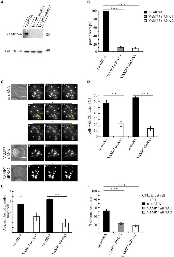
Knockdown of VAMP7 strongly reduces fusion of cytotoxic granules at the IS. (A) Lysates from bead stimulated human CD8+ T cells transfected with either control or VAMP7 siRNAs (1 or 2, respectively) and blotted for VAMP7 (top) and GAPDH (bottom) as loading control. (B) Quantification of VAMP7 protein expression (in % normalized to control siRNA-treated CTLs) performed by densitometry. Bars indicate SEMs. [VAMP7-siRNA1, N = 3; ***p < 0.001 and VAMP7-siRNA2, N = 3; ***p < 0.001 (t-test)]. (C) Human CD8+ T cells co-transfected with granzyme B-mCherry along with either ns-siRNA or VAMP7-siRNA1 or VAMP7-siRNA2 and imaged 12 h after transfection. Selected live-cell TIRF microscopy images of granzyme B-mCherry in a transfected CTL in contact with an anti-CD3 coated coverslip. Fusion events are indicated with open circles (three frames shown per granule fused). (D) Mean percentage of cytotoxic granule fusion in cells transfected with either ns-siRNA (n = 66 and n = 59, respectively) or VAMP7-siRNA1 [n = 91; **p < 0.01 (t-test)] or VAMP7-siRNA2 [n = 72; ***p < 0.001 (t-test)]. (E) Mean average number of granules fused over time in the TIRF plane per cell p = 0.206 (t-test) for VAMP7-siRNA1 and **p < 0.01 (t-test) for VAMP7-siRNA2. Bars indicate mean ± SEM. Scale bar, 5 μm. (F) Calcein-based killing assay for CTLs transfected with either ns-siRNA, VAMP7-siRNA1, or VAMP7-siRNA2. Experiments were carried out in duplicate [N = 4; ***p < 0.001 (t-test)].

