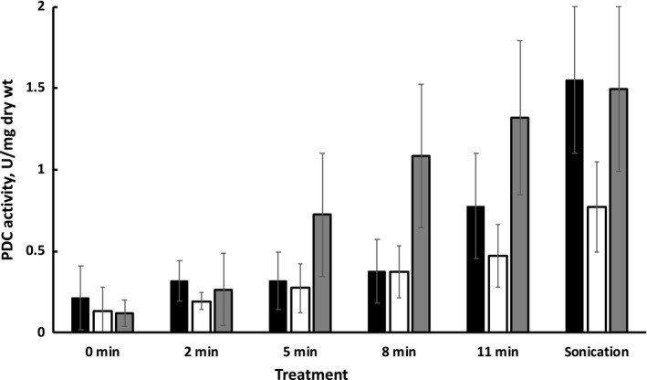
Figure 3.

PDC activity in cell‐free extracts and in extracellular medium after lysozyme treatment of various duration: Zm6 (black columns), Pdc‐ (empty columns), and PeriAc (gray columns). Assay for lysozyme treatment: 100 L of 100 mg/ml lysozyme was added to 900 L of 4 mg/ml washed cell suspension (harvested at mid‐exponential phase of aerobic growth) in 0.2 M TRIS/HCl buffer, pH 8, and incubated at room temperature by vortexing of various duration. Then, samples were rapidly cooled in an ice bath, centrifuged, and the supernatants used for PDC activity measurement. Cell‐free extracts were prepared by 2 min ultrasonic disruption with pulses of 0.5 s duration, separated by 0.5 s intervals. PDC activity assay: 5 L of the supernatant from lysozyme‐treated or sonicated cell suspension was added to a mixture, containing 937 L of 0.2 M citrate buffer, pH 6, 38 L of 1 M pyruvate, 15 L of 10 mM NADH, and 5 L of ADH. Results represent mean values of five experiments, with error bars showing standard deviation
