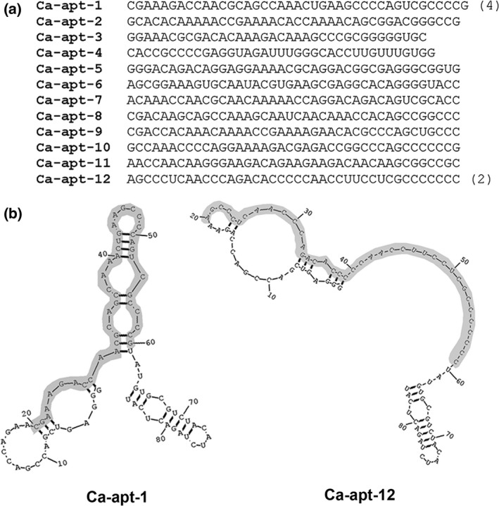
Figure 1.

The sequences and predicted secondary structure of the Candida albicans‐specific aptamers (Ca‐apt). (a) The randomized region of the aptamers is shown. The number in parentheses represents the number of clones identified during the aptamer screening. (b) The predicted secondary structures of Ca‐apt‐1 and Ca‐apt‐12 with the lowest folding energy are shown. The nucleotides in the shaded area correspond to the randomized region of the aptamer
