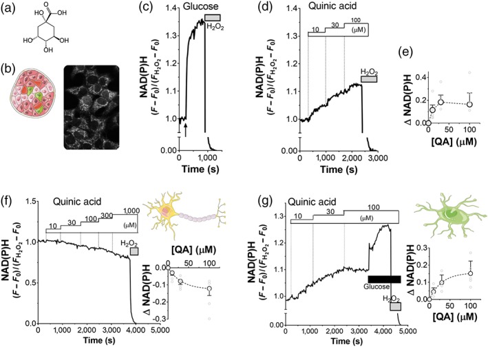
Figure 1.

Quinic acid (QA) enhances oxidative metabolism in INS‐1E cells and iCell astrocytes but not in iCell neurons. (a) QA (structure) enhances oxidative metabolism in INS‐1E cells by stimulating Ca2+‐dependent mitochondrial dehydrogenases, which produce NADH. (b) NAD(P)H autofluorescence in INS‐1E cells at 2.5‐mM glucose. Representative traces of NAD(P)H autofluorescence of INS‐1E cells, stimulated with (c) 16.7‐mM glucose and (d) QA (dose–response, as indicated). (c–g) The NAD(P)H signal was normalized to the fluorescence measured in the basal Krebs–Ringer bicarbonate HEPES buffer (set to 1) minus the minimal signal after full oxidation to NAD(P)H using 1% H2O2 (set to 0). Data are representative of (c) n = 5 and (d) n = 5 experiments. (e) Dose–response effect of QA on mitochondrial NAD(P)H production. Values shown are means ± SEM from n = 5 experiments. Effect of QA on NAD(P)H production in (f) iCell neurons and (g) iCell astrocytes. Representative traces (N = 5) of NAD(P)H autofluorescence, stimulated with QA, at the indicated concentrations. Normalization as described in (c). (g) Glucose (2mM) was added, as indicated. Dose–response amplitude of NAD(P)H response, evoked by QA in (f) iCell neurons and (g) iCell astrocytes. Values shown are means ± SEM from n = 5 experiments
