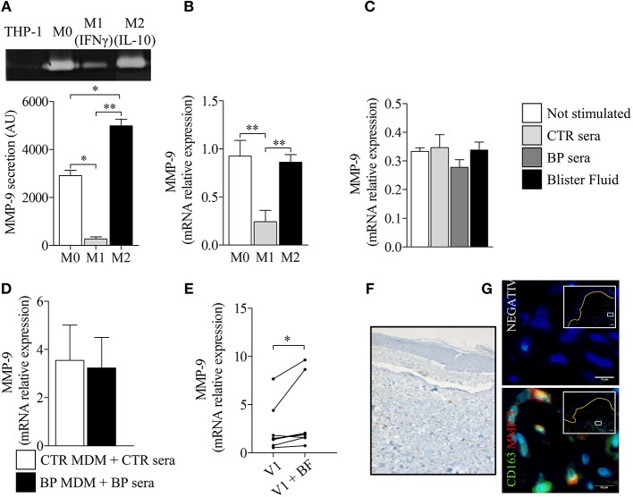
Figure 6.
Polarization of BP-MdM influenced their capacity to produce the protease MMP-9. MMP-9 secretion (A) and MMP-9 mRNA expression (B) were analyzed, respectively, by gelatin zymography and real-time qPCR in M0, M1 (+IFNγ), and M2 (+IL-10) THP-1 derived macrophages. The error bars denote the mean ± SEM. Paired Student's T-Test was used for statistics (n = 3; *p < 0.05; **p < 0.01). (C–E) MMP-9 mRNA expression was analyzed by real-time qPCR in both THP-derived (C) and BP monocyte-derived macrophages (D,E) stimulated with either control serum, BP serum or blister fluid from BP patient. The error bars denote the mean ± SEM. Nonparametric unpaired Mann-Whitney's test was used for statistical analysis of (C,D) (n = 8 and n = 5, respectively). Nonparametric paired Wilcoxon's test was used to statically analyze (E) (n = 8; *p < 0.05). (F,G) Punch biopsies of lesional skin from patients with BP were subjected to CD163 immuno-staining alone (F) or double immunofluorescence staining for MMP-9 (red) and CD163 (green) (G) with Hoescht counterstain (blue). Negative CTR, negative control where primary antibodies were not added. White islets show lower magnification images of blister area and orange line designed the dermo-epidermal junction. The boxes in white islets match to the area where the high magnification image has been taken.

