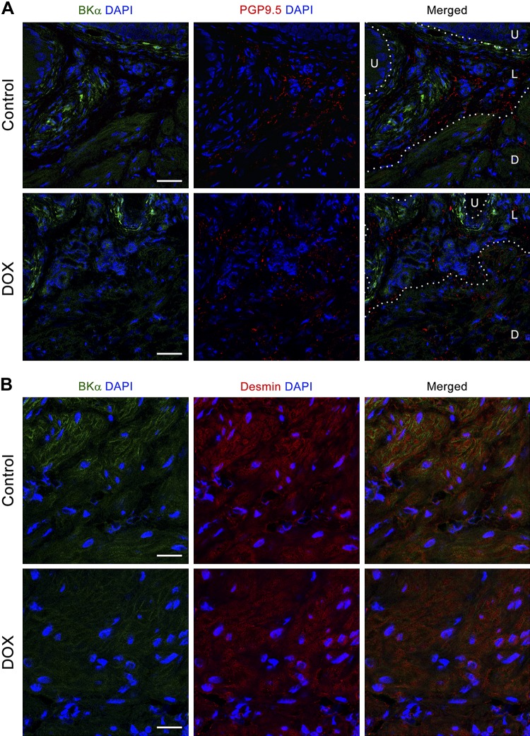
Fig. 6.
Distribution of the large-conductance Ca2+-activated K+channel α-subunit (BKα) in the bladders. A: representative immunostainings for BKα (green; left), protein gene product 9.5 (PGP9.5; red; middle), and merged image (right) with DAPI (blue) for nuclear staining. White, dotted lines outline the boundaries among the urothelial (U), lamina propria (L), and detrusor smooth muscle (D) layers. Original scale bars = 50 µm. B: representative immunostainings for BKα (green; left), desmin (red; middle), and merged image (right) with DAPI (blue) for nuclear staining (N = 4 per group). Original scale bars = 10 µm. DOX, doxorubicin.

