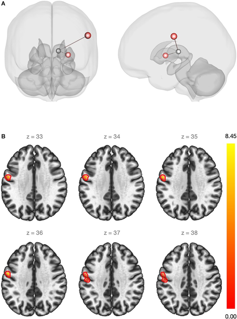
FIGURE 1.

(A) Red regions show an increase in the rs-FC between the left thalamus and the left sensorimotor cortex (beta = 0.20)/left putamen (beta = 0.14) after estradiol therapy (ROI-to-ROI analysis). Statistical significance thresholded at p-FDR <0.05. (B) Whole-brain seed-to-voxel functional connectivity analysis. Red cluster indicates increased rs-FC between the left thalamus (seed) and voxels of the pre and post-central gyri (beta = 0.21). Cluster size p-FDR <0.0042. Color bar shows statistical significance.
