Figure 5.
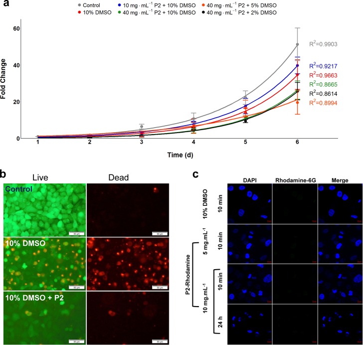
Post-thaw growth and polymer interaction. (a) Post-thaw cell proliferation for 6 d; cells were seeded at 1.25 × 10⁴ cells per well. (b) LIVE/DEAD staining of A549 cells; 24 h post-thaw after cryopreservation using indicated cryoprotectants. The scale bar is 50 μm. (c) Confocal fluorescence microscopy of A549 cells incubated with 10% DMSO or P2 (with no DMSO) for the indicated time and concentration. Nuclei are stained with 4′,6-diamidino-2-phenylindole) (DAPI) and incubated with P2 tagged with rhodamine 6G, which is green fluorescent. The scale bar is 10 μm.

