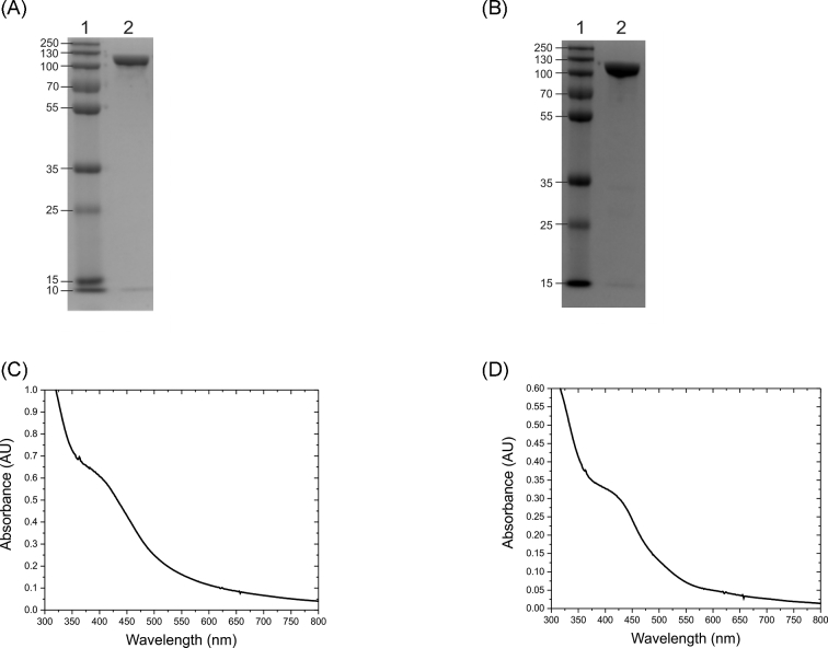
Figure 1.
Characterization of native and variant C. jejuni NapA by gel electrophoresis and UV-vis spectroscopy. (A) SDS PAGE image of the NapA, (B) SDS PAGE image of the NapA-C176S variant where lane 1 is the molecular weight standards in kDa, (C) UV-vis spectrum of the as-prepared recombinant NapA in 50 mM HEPES pH 7.00, (D) UV-Vis spectrum of the as-prepared NapA-C176S variant in 50 mM HEPES pH 7.00.

