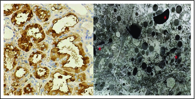
Figure 4.
Features of LCPT. IHC stained strongly positive for κ light chain in proximal tubules; λ (data not shown) was negative (original magnification ×400; DAB + Harris’s hematoxylin stain; left panel). TE microscopy image shows rhomboid crystal inclusions (*) in keeping with light chain proximal tubulopathy (original magnification ×4000; right panel).

