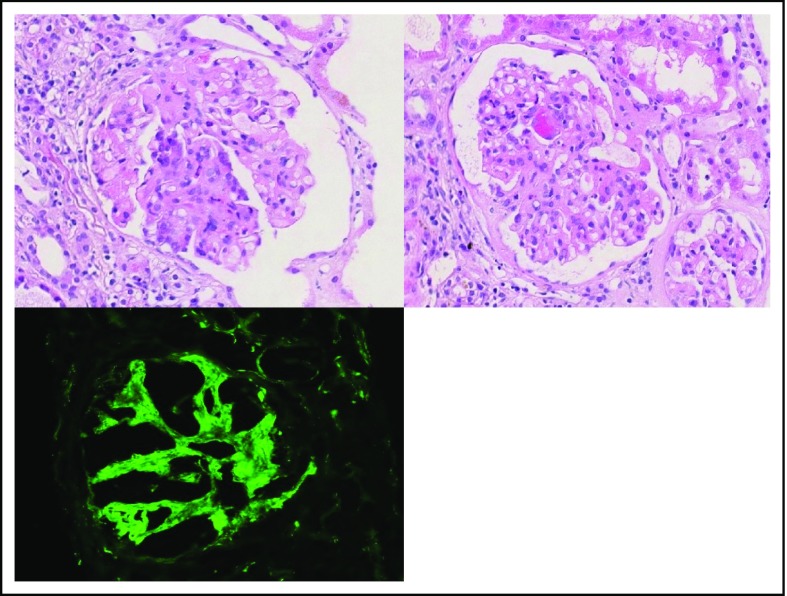
Figure 6.
Features of a C3GN in the setting of MGRS. Glomerulus with segmental endocapillary hypercellularity (original magnification ×400; hematoxylin & eosin stain; upper left panel). Glomerulus with segmental capillary tuft fibrinoid necrosis (original magnification ×400; hematoxylin & eosin stain; right panel). IF showed C3-dominant deposits in the mesangium and capillary loops, in keeping with a C3-dominant GN (original magnification ×400; fluorescein isothiocyanate; lower left panel).

