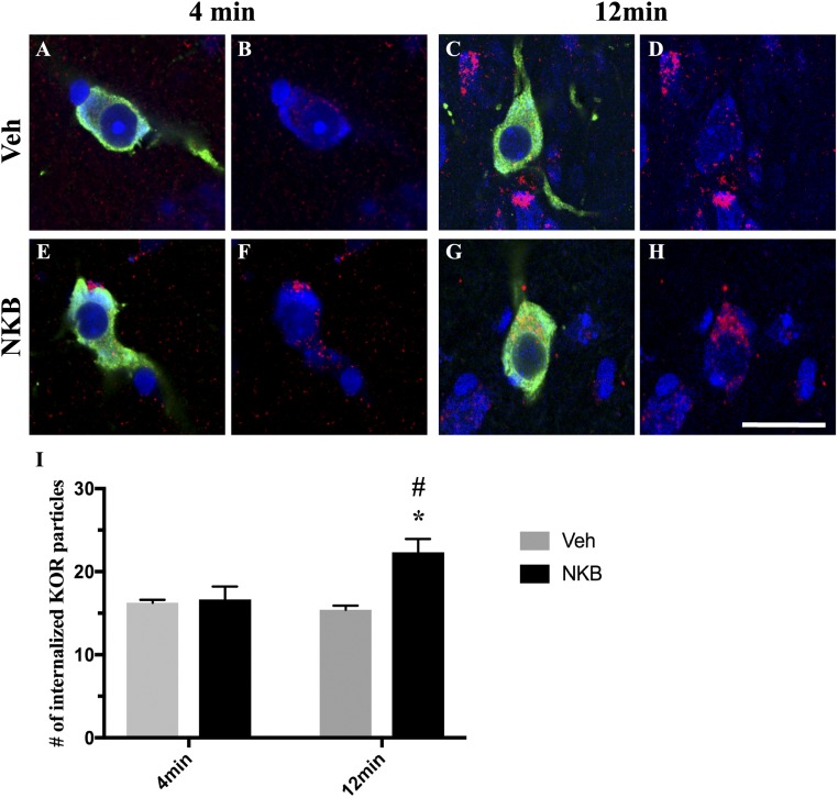
Figure 5.
Confocal images of 1-μm-thick optical sections showing internalized KOR (red) particles in MBH GnRH (green) neurons counterstained with fluorescent Nissl (blue) (A and E) 4 min and (C and G) 12 min after (A and C) Veh or (E and G) NKB injection. (B, D, F, and H) Same images of GnRH cells as in panels (A), (C), (E), and (G) but showing only KOR (red) and fluorescent Nissl (blue) to enhance visualization of internalized particles. Scale bar, 20 μm. (I) Mean numbers (±SEM) of internalized KOR particles in GnRH-positive cells in the MBH in ewes 4 min and 12 min postinjection of Veh (gray bars) and NKB (black bars). *P < 0.05, Veh vs NKB within time points; #P < 0.05, 4 min vs 12 min within treatment group.

