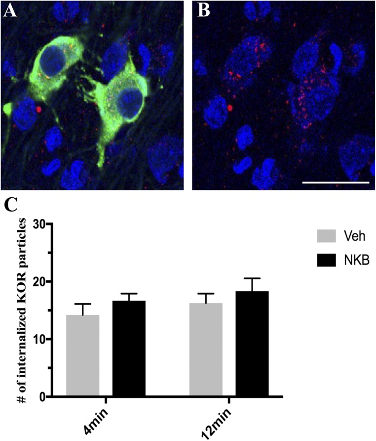
Figure 6.
(A) Representative confocal image of 1-μm-thick optical sections showing internalized KOR (red) particles in GnRH (green) neurons within the POA counterstained with Fluoro Nissl (blue) 12 min post NKB injection. (B) Same image of GnRH cells as in (A) but showing only KOR (red) and fluorescent Nissl (blue) to enhance visualization of internalized particles. Scale bar, 20 μm. (C) Mean numbers (±SEM) of internalized KOR particles in GnRH-positive cells in the POA in ewes 4 min and 12 min postinjection of Veh (gray bars) and NKB (black bars).

