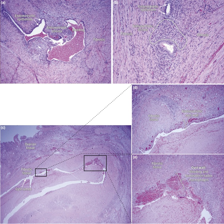
Figure 4.
(a) Peritoneal endometriosis with fibrosis. (b) Rectovaginal nodule with extensive fibrosis and tissue remodeling surrounding islands of endometriotic stroma and occasional epithelial cells. (c) Sections of an ovarian endometrioma cyst. Note that the thickness of the endometrial lining (stroma and luminal epithelium) varies throughout the cyst wall, with foci of bleeding and macrophages containing blood pigment. The cyst wall is primarily composed of fibrotic tissue. (d and e) Higher-magnification pictures showing details of the ovarian endometrioma cyst wall from (c).

