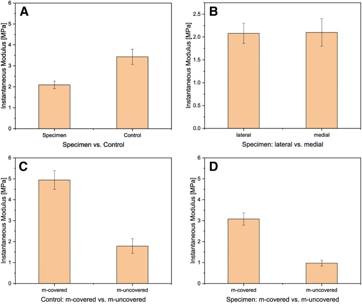
Fig. 2.
Comparison of instantaneous modulus for a specimen and control: specimen N = 25, control N = 13; p < 0.05. b Comparison of the IM and its location: medial vs lateral; medial N = 21, lateral N = 25. Comparison of the IM and its location: c cartilage covered vs not covered by meniscus (for medial and lateral); d specimen, m-covered N = 31, m-uncovered N = 30; right: controls, m-covered N = 25, m-uncovered N = 23

