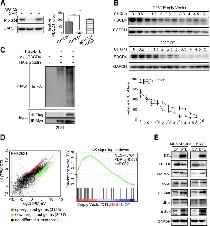
Fig. 4.
DTL degraded PDCD4 and activated JNK pathway. a 293 T cells treated with 50 mg/mL cycloheximide alone or plus 10 mmol/L MG132 for 6 h. PDCD4 expression was detected. b 293 T cells were transient transfected by DTL or empty vector and treated with 50 mg/mL cycloheximide (CHX) for indicated times. The diagram showed quantitative analysis of PDCD4 levels. c HA-ubiquitin and Myc-PDCD4 were co-transfected into 293 T cells with or without DTL. Immunoprecipitation with anti-Myc antibody followed by Western blot using HA antibodies showed DTL promotes PDCD4 ubiquitination level. d RNA sequencing was performed in 293 T cells with empty vector and DTL respectively. Pathway enrichment analysis of differential expression genes revealed that JNK pathway was the most enriched. e Western blot was used to confirm the results obtained by RNA sequencing in cancer cells. *, P < 0.05, **, P < 0.01 based on the Student t test. All results are from three or four independent experiments. Error bars, SD

