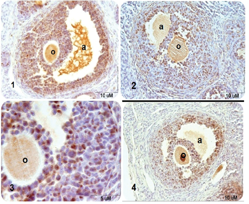
Figure 1.

Microscopic sections of rat's secondary ovarian follicles after PCNA immunohistochemical technique. PCNA-positive nuclei are seen in brown- (1) control group, (2) DZN-treated group in which DZN decreased the PCNA-positive cells significantly compared to the control group, (3) DZN-treated group by magnification 100, and (4) DZN+vitamin E-treated group in which the number of PCNA-positive cells increased significantly compared to the DZN group. Note: a: antrum; o: oocyte.
