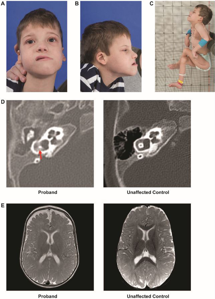
Figure 1. Clinical phenotype.

A. A photo of the proband depicting an oblong face with a long lower-third face, forward placed midface, and bitemporal and bicoronal narrowing.
B. Side profile of the proband showing long lower-third face, forward placed midface, and low-set ear with curling of the pinna
C. Typical posture and positioning of proband. Although hypotonic, he has significant contractures. Scar visible from osteotomy.
D. Computed tomography of the temporal bone showing vestibular dysplasia (left) as compared with an unaffected 5-month old.
E. Magnetic resonance imaging of the brain, with axial section, showing decreased cerebral volume and enlarged arachnoid spaces (left) as compared with an unaffected 9-month old.
