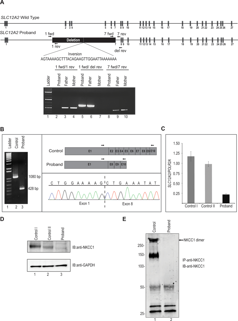
Figure 2. Molecular analysis of SLC12A2 in proband.

A. Upper, schematic representation of wild type SLC12A2 locus. Middle, structure of proband’s SLC12A2 locus showing homozygous deletion of 22kb and inversion of 34 nucleotides. Arrows indicate position of forward and reverse primers used for PCR amplification. Lower, agarose gel image showing amplification of genomic DNA demonstrates homozygous deletion in proband and heterozygous deletion in his father. The proband’s genomic DNA from blood amplifies only when primers are used outside of the deletion. The father’s DNA amplifies in all cases, showing he is a heterozygote. The mother’s genomic DNA does not amplify when primers straddle the breakpoint, as this region is presumably of a restrictive, wild type length of 22kb.
B. Analysis of cDNA synthesized from total RNA extracted from control and proband’s dermal fibroblasts. PCR amplification of cDNA results in truncated transcript in proband. Lane 1 is 1Kb molecular weight DNA ladder. Arrows indicate position of forward and reverse primers used for cDNA amplification. Cloning of PCR product followed by sequencing showed direct splicing of exon 1 to downstream exon 8 in proband.
C. Quantitative real time PCR analysis revealed significant reduced expression of SLC12A2 transcript normalized to POLR2A in proband fibroblasts compared to two independent fibroblast controls.
D. Western blot analysis of protein lysates from fibroblasts probed with polyclonal anti-SLC12A2 (NKCC1) antibody shows no detectable SLC12A2 protein in proband compared to two independent controls. The lower panel shows western blotting with anti-GAPDH to confirm equivalent sample loading.
E. Protein lysates from fibroblasts were immunoprecipitated with polyclonal anti-SLC12A2 (NKCC1) antibody conjugated to Protein A sepharose. Western blotting of immunoprecipitates with monoclonal anti-SLC12A2 (NKCC1) revealed the presence of SLC12A2 in both monomeric and dimeric forms in control while no SLC12A2 bands were detected in proband. Molecular weight in kilodaltons (kDa) is shown to the left of the panel.
