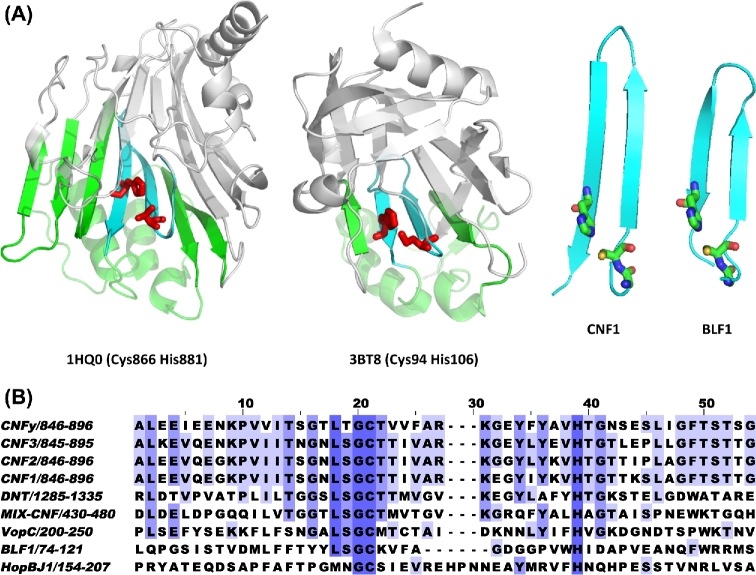
Figure 3.
CNF1 signature active-site motif. (A) Shown are PyMOL renderings of the crystal structures of the C-terminal CNF1 catalytic domain (1HQ0) and the Burkholderia lethal factor 1 (3BT8) (right panels). The bottom portion of the structure (green) contains the beta-turn-beta motif (cyan) with the active site His and Cys residues shown in red. The portion of the structure above the active site beta sheet is shown in grey. Shown to the right are the corresponding zoomed-in views of the beta-turn-beta motifs with Gly, Cys and His residues shown as stick structures. (B) Alignment of sequences in the region near the Gly-Cys-beta-turn-beta-His motif in proteins known to have CNF1-like Gln-deamidase activity.

