Figure 1.
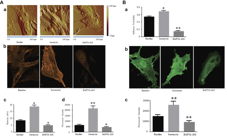
(A) [Ca2+]i regulates Expression of α-SMA and cellular stiffness in VSMCs. Ionomycin increased [Ca2+]i, but BAPTA-AM decreased [Ca2+]i. (a) Atomic force microscopy topography analysis of VSMCs treated with or without drugs by increasing or decreasing [Ca2+]i. The VSMC area and height increase by a higher [Ca2+]i level, and those decrease by a lower [Ca2+]i level. (b) Atomic force microscopy stiffness analysis of VSMCs treated with or without drugs by increasing or decreasing [Ca2+]i. (c) Representative confocal immunofluorescent images showed α-SMA expression of VSMCs treated with or without drugs by increasing or decreasing [Ca2+]i. (d) α-SMA-positive signals among VSMCs treated with or without drugs by increasing or decreasing [Ca2+]i. Results are expressed as mean ± SEM, **p < 0.01 and *p < 0.05 vs. baseline. Reprinted from Zhu et al. (2018b) by published permission. (B) [Ca2+]i regulates α5 integrin subunit expression and α5β1 integrin adhesion activities in VSMCs. Ionomycin increased [Ca2+]i, but BAPTA-AM decreased [Ca2+]i. (a) Atomic force microscopy measurement of probability of adhesion to FN matrix of VSMCs treated with or without drugs by increasing or decreasing [Ca2+]i. (b) Representative confocal immunofluorescent images showed α5 integrin expression of VSMCs treated with or without drugs by increasing or decreasing [Ca2+]i. (c) α5 integrin-positive signals among VSMCs treated with or without drugs by increasing or decreasing [Ca2+]i. Results are expressed as mean ± SEM, **p < 0.01 and *p < 0.05 vs. baseline. Reprinted from Zhu et al. (2018b) by published permission.

