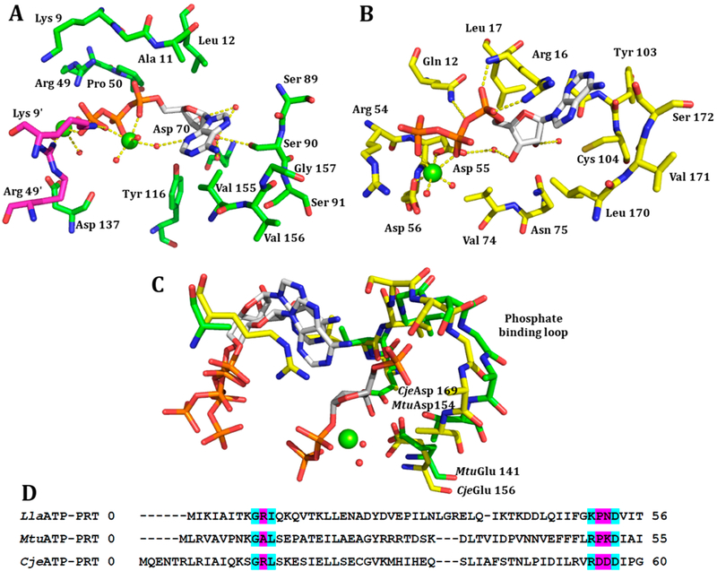
Figure 4.
(A) ATP bound to the active site of CjeATP-PRT (PDB: 4YB7) with the main chain in yellow and H-bonds as yellow dashed lines. (B) ATP bound to the active site of MtuATP-PRT (PDB: 5U99) with the main chain in green and second chain in purple, H-bonds as yellow dashed lines. (C) Overlay of the conserved phosphate binding loop between PRPP bound Giardia lamblia adenine PRT (PDB: 1L1R), ATP bound CjeATP-PRT (PDB: 4YB7), and ATP bound MtuATP-PRT (PDB: 5U99). (D) A partial sequence alignment with pink highlighted regions showing low levels of conservation between the three enzymes leading to changes in the binding mode of ATP. Cyan highlighted residues show high levels of conservation.

