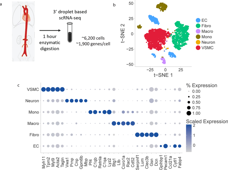
Figure 1:
Single cell RNA-sequencing atlas of aortic cells types. a) Schematic overview of experimental approach. Four mouse aortas were dissected from aortic root to femoral take-off, enzymatically dissociated for 1 hour at 37ºC, and then the single cell suspension was sequenced at a depth of 1,900 median genes per cell using droplet-based RNA-sequencing methods. b) t-SNE representation of single cell gene expression shows the 7 identified major aortic cell types. c) Dotplot demonstrates the top markers of each aortic cell type. Dot size corresponds to proportion of cells within the group expressing each transcript and dot color corresponds to expression level. d) Heatmap identifying all genes with log fold change>2 for each aortic cell type relative to all other cells. EC indicates endothelial cells; Fibro, fibroblasts; Macro, macrophages; Mono, monocytes; VSMC, vascular smooth muscle cells.


