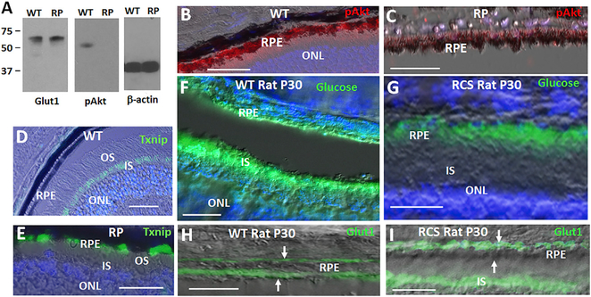
Figure 2. A Tam/pAkt/Txnip/Glut1 Pathway in RPE Glucose Transport.
(A) Western blot of RPE from WT and RP mice at P40. pAkt = pAktS473.
(B and C) pAkt in RPE from WT mice (B) is diminished in the cells in RP (C). Immunostaining for Akt phosphorylated on S473 (pAktS473) is shown at P40.
(D and E) Txnip is absent in the RPE of WT mice (D), but it induced in the cells in RP (E). Txnip in photoreceptor inner segments (IS) of WT mice (D) is downregulated in IS in RP mice at P40 (E).
(F) Fluorescently labeled 2-deoxyglucose (“glucose”) transport to photoreceptor IS in WT rats at P30.
(G) Diminished transport of glucose to photoreceptors in RCS rat littermates at P30.
(H and I) Diminished Glut1 expression on the RPE apical surface in RCS rats compare to WT littermates at P30. (H) and (I) show immunostaining for Glut1. Arrows show the basal and apical surfaces of the RPE. Bars are 50 μm.

