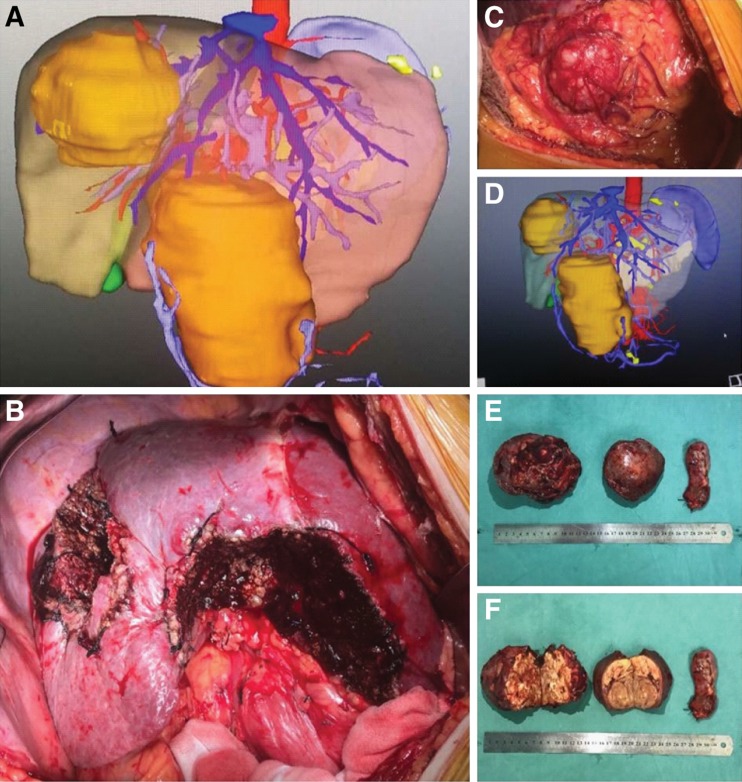
Figure 2.
A representative set of the three‐dimensional computed tomography (CT) imaging and operative photographs of a 62‐year‐old male with symmetrical HCCs located in bilateral hemilivers. Enhanced CT three‐dimensional imaging (A, D) of a 62‐year‐old man shows two large lesions located in Segments 7 and 8 (right liver, tumor size: 12.0 cm) and Segments 3 and 4 (left liver, tumor size: 12.5 cm), respectively. His preoperative alpha‐fetoprotein level was 825 ng/mL (normal value: ≤20 ng/mL). This patient underwent curative liver resection for symmetrical large binodular hepatocellular carcinomas (B, C, E, F) on October 27, 2015, and was alive and recurrence‐free on the last date of follow‐up on December 1, 2018.

