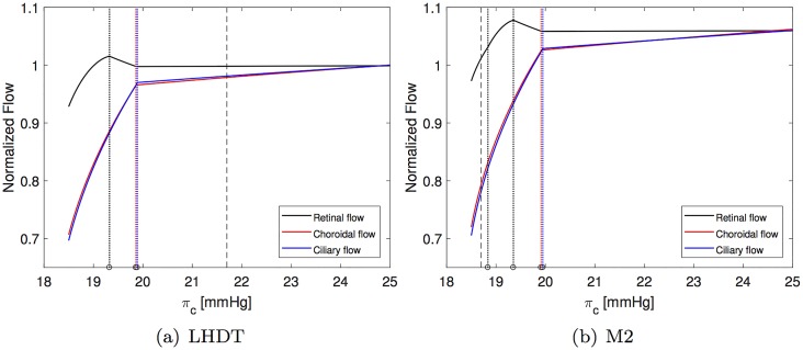
Fig 5. Normalized fluxes in the retina, choroid and ciliary body as a function of the blood oncotic pressure in capillaries πc.
All fluxes are normalized with their respective baseline value. The vertical dashed line indicates the value of πc representative of each case. Dotted colored vertical lines indicate the values of πc at which each of compartment collapses (black for central retinal vein, red for choroidal venules and blue for ciliary venules). Panel (b) appeared in [12].

