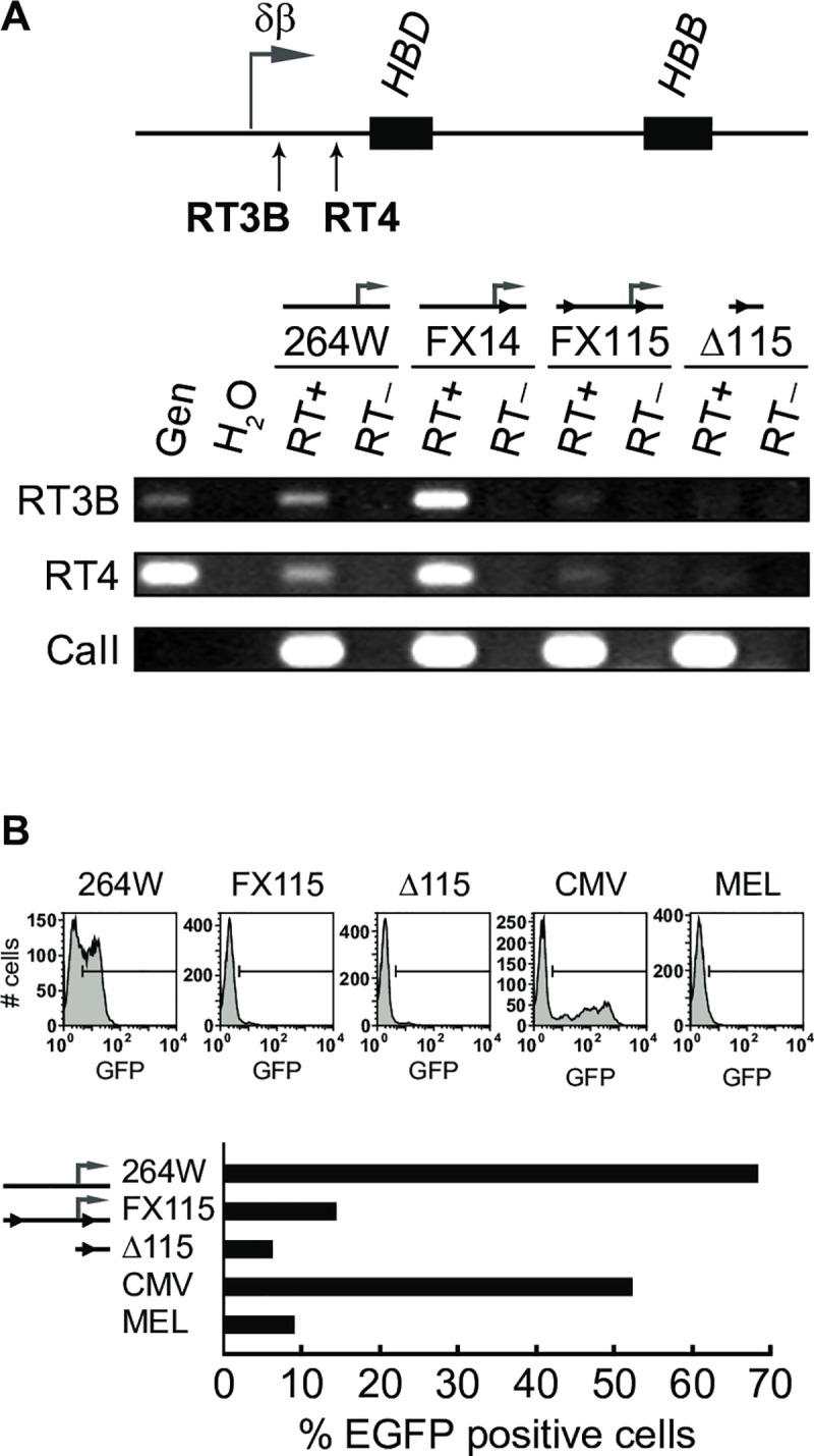
Fig 5. Intergenic transcription in the β-globin locus before and after deletion of the δβ intergenic promoter.

(A) RT-PCR to detect intergenic transcripts downstream of the δβ intergenic promoter in adult erythroid cells. The positions of PCR amplicons RT3B and RT4 are indicated below the map; black boxes, HBD and HBB genes; horizontal arrow, normal position of the δβ intergenic promoter and transcript start site. Intergenic transcription is reduced in lines FX115 and Δ115 with ‘floxed’ and deleted δβ intergenic promoter, respectively. (B) Transfection assays to assess intergenic promoter activity. Fragments containing the δβ intergenic promoter or corresponding fragments from the deletion lines were PCR amplified from lines 264W, FX115, and Δ115, and cloned upstream of a promoterless EGFP gene as described in Materials and Methods. Reporter constructs were stably transfected into MEL cells and the number of EGFP-expressing cells in the population was determined by flow cytometry. The percentage of cells expressing EGFP is severely decreased compared to wild-type δβ promoter (assigned an arbitrary value of 100) and CMV promoter when driven by FX115- and Δ115-derived sequences.
