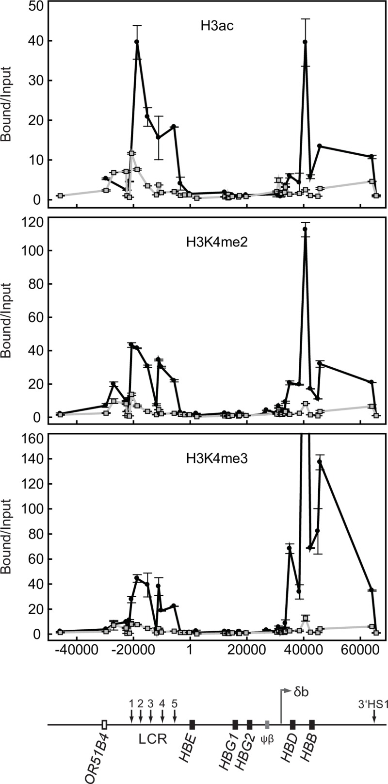
Fig 7. Histone modifications across the human β-globin locus in wt and δβ promoter deleted adult erythroid cells.

Chromatin immunoprecipitation assays for histone H3 acetylation (H3ac), di-methylated lysine 4 of histone H3 (H3K4me2), and tri-methylated lysine 4 of histone H3 (H3K4me3); ●, wild-type adult erythroid cells from line 264W [10];  , adult erythroid cells from line Δ115. Coordinates in bp are shown along the x axis with HBE start site as +1. Bound versus input ratios were calculated and normalized to the most 3’ primer pair, located just downstream of 3’HS1 in the ORG cluster. A map of the locus is shown below the graphs.
