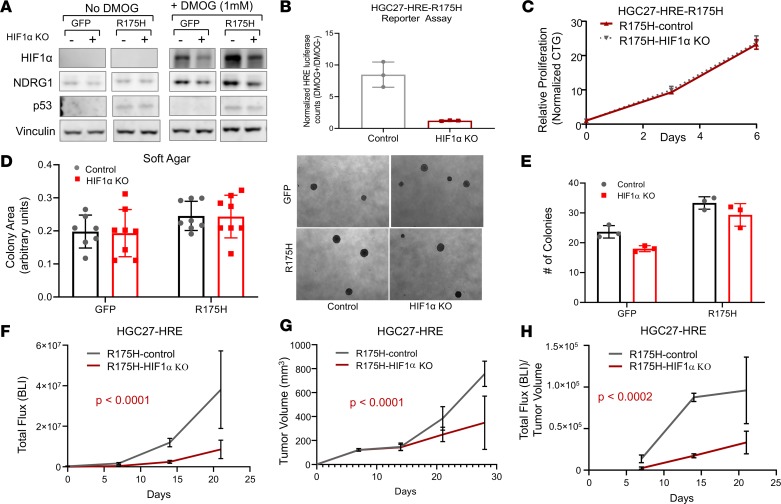
Figure 7. Disruption of HIF1α impairs primary tumor growth of mutant p53 gastric cancer.
(A) Immunoblot of HIF1α and NDRG1 expression levels in HGC27-HRE cells stably expressing either control or HIF1α sgRNA in the presence and absence of 1 mM DMOG. (B) Luciferase assay of HGC27-HRE-R175H cells stably expressing either control or HIF1α sgRNA displayed as a ratio of 1 mM DMOG/no DMOG. (C) Proliferation of HGC27-HRE-R175H cells stably expressing either control or HIF1α sgRNA under adherent culture conditions as measured by CTG; normalized to baseline counts on day 0. (D and E) Soft agar growth of HGC27-HRE-GFP and HGC27-HRE-R175H cells stably expressing either control or HIF1α sgRNA. Right panel in D shows representative images of colonies. (F) Total HRE flux (bioluminescence) of HGC27-HRE-R175H–expressing control (n = 5) or HIF1α sgRNA (n = 5) xenografts; comparison of fits, P < 0.0001. (G) Tumor volume of HGC27-HRE-R175H–expressing control (n = 5) or HIF1α sgRNA (n = 5) xenografts; comparison of fits, P < 0.0032. (H) Total HRE flux/tumor volume of HGC27-HRE-R175H–expressing control (n = 5) or HIF1α sgRNA (n = 5) xenografts; comparison of fits, P < 0.0002. All data are presented as mean ± SD.

