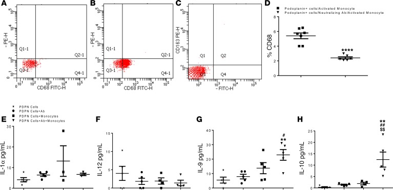
Figure 7. Podoplanin neutralization represses monocyte activation in vitro.
Flow-cytometry analysis of cultured podoplanin positive cells and bone marrow isolated monocytes cultured in vitro and stimulated with LPS. (A) Podoplanin positive cells do not express CD68, (B) LPS activated monocyte highly expressed CD68 and were negative for CD163 (C). The flow-cytometry scatterplots are representive of biological triplicate isolations. (D) The graph represents the quantification of the flow-cytometry analysis of the monocytes CD68 expression after cocultured with podoplanin positive cells, with and without the presence of podoplanin neutralizing antibody. Data shows significant reduction of CD68 expression when podoplanin positive cells and LPS-stimulated monocyte were co-cultured in the presence of podoplanin neutralizing antibody. Data are presented as mean ± SEM ****P < 0.01 monocytes cocultured with podoplanin positive cells vs. monocytes co-cultured with podoplanin positive cells in the presence of podoplanin neutralizing Ab. Student’s t-test was performed between the groups. n = 3/group, the data from biological triplicates in each separate experiment were consistent. Conditioned medium obtained from podoplanin (PDPN) positive cells only, podoplanin positive cells treated with neutralizing podoplanin antibody, podoplanin positive cells cocultured with activated monocytes with and without the presence of neutralizing podoplanin antibody were analyzed by ELISA for proinflammatory (E and F) and antiinflammatory (G and H) cytokines. The presence of the neutralizing antibody drastically reduced secretion of IL-1α (E) and increased IL-10 secretion (H). Data are presented as mean ± SEM **P < 0.02 podoplanin positive cells only vs. podoplanin positive cells co-cultured with activated monocytes with neutralizing antibody, #P < 0.05 and ##P < 0.02 podoplanin positive cells with podoplanin neutralizing antibody vs. podoplanin positive cells cocultured with activated monocytes with neutralizing antibody, $$P < 0.02 podoplanin positive cells cocultured with activated monocytes without neutralizing antibody vs. podoplanin positive cells cocultured with activated monocytes with neutralizing antibody. n = 3–5/group. One-way ANOVA analysis and Bonferroni post hoc test have been performed among all groups.

