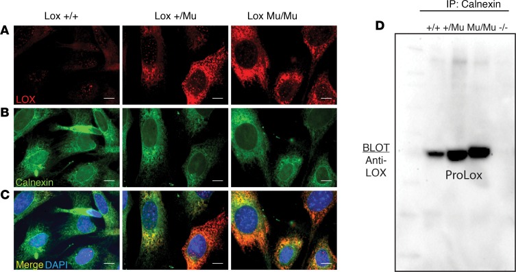
Figure 5. Intracellular mutant LOX interacts with ER-resident protein calnexin.
(A) Immunofluorescence imaging of Lox+/+, Lox+/Mu, and LoxMu/Mu mouse embryonic fibroblasts (MEFs) using an anti-LOX antibody showed intracellular accumulation of mutant but not WT LOX protein. (B) Imaging using an antibody to calnexin showed a uniform distribution of intracellular calnexin staining in all cells. (C) Merged images of LOX and calnexin staining established colocalization of intracellular mutant LOX with calnexin. Scale bars: 1 μm (D) Cell lysates from Lox+/+, Lox+/Mu, LoxMu/Mu, and Lox–/– MEFs were immunoprecipitated with anti-calnexin antibody, separated by SDS-PAGE, then immunoblotted with an anti-LOX antibody. Both WT and mutant LOX were immunoprecipitated with calnexin, but the level of calnexin-bound mutant LOX was much higher than that of WT LOX. Cell lysate from Lox–/– cells served as a negative control.

