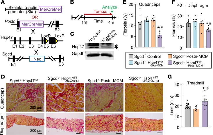
Figure 4. Myofibroblast-specific but not myofiber-specific deletion of Hsp47 in skeletal muscle reduces muscular dystrophy–dependent tissue fibrosis.
(A) Schematic of the MCM cDNA driven by the human skeletal α-actin promoter (myofiber-specific) or the myofibroblast-specific Postn genetic locus to delete the Hsp47 gene with tamoxifen treatment. These lines were crossed into the δ-sarcoglycan–null (Sgcd–/–) background. (B) Experimental tamoxifen dosing regimen administered in the feed. (C) Western blot analysis for total Hsp47 protein using whole muscle protein lysates from the quadriceps of mice of the indicated genotypes. n = 6 mice per group. Gapdh is shown as a loading control. (D) Representative Picrosirius red–stained histological sections from quadriceps and diaphragm from 4-month-old mice of the indicated genotypes. Scale bar: 200 μm. (E and F) Quantitation of fibrosis from Picrosirius red–stained histological sections from quadriceps and diaphragm of 4-month-old mice of the indicated genotypes. n = 7 mice in each group. (G) Average time spent running on a treadmill of 4-month-old mice of the indicated genotypes. n = 6–10 in each group. Significance was determined using P values calculated by 1-way ANOVA with Tukey’s post hoc test. *P < 0.05 versus Sgcd–/–-control. #P < 0.05 versus Sgcd–/– Hsp47fl/fl-Ska-MCM. The legend applies to E, F and G.

