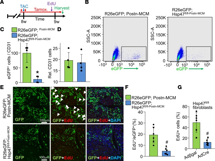
Figure 6. Myofibroblast-specific Hsp47 deletion reduces myofibroblasts in vivo and their proliferation.
(A) Experimental scheme whereby mice were subjected to TAC injury for 7 days. Mice received 2 i.p injections of tamoxifen and were fed tamoxifen-laden chow 48 hours before surgery and were then maintained on this chow until harvesting. Mice also received a single i.p EdU injection 4 hours before sacrifice at day 7 after TAC. (B) Representative flow cytometry plots of isolated EGFP+ interstitial cells (plotted as EGFP fluorescence signal on the x axis versus side scatter on the y axis) from hearts of the indicated genotypes of mice; 100,000 cells are displayed in the blots. (C) The ratio of total EGFP+ myofibroblasts normalized to CD31+ cells from the hearts of the indicated genotypes of mice after 1 week of TAC. Error bars represent SEM; n = 4 mice in each group. *P < 0.05 versus Postn-MCM; R26eGFP. P values were calculated with a Student’s t test. (D) Relative number of CD31+ cells in the interstitial fractions in hearts of the indicated genotypes of mice after 1 week of TAC. (E) Representative immunohistological images (scale bar: 100 μm) of EdU+ and EGFP+ interstitial cells at the time of harvest for mice treated, as shown in A. DAPI was used to show nuclei (blue). n = 5 mice in each group. (F) Quantitation of GFP+ cells that were also EdU+ in heart histological sections from mice subjected to TAC of the indicated genotype. P values were calculated with Student’s t test. #P < 0.05 versus R26eGFP Postn-MCM controls. (G) Quantitation of EdU+ Hsp47fl/fl cardiac fibroblasts over 24 hours in culture previously treated with AdCre or Adβgal infection. A total of 6 images were analyzed per group. P values were calculated with Student’s t test. #P < 0.05 versus Adβgal infection. Data shown are the mean ± SEM.

