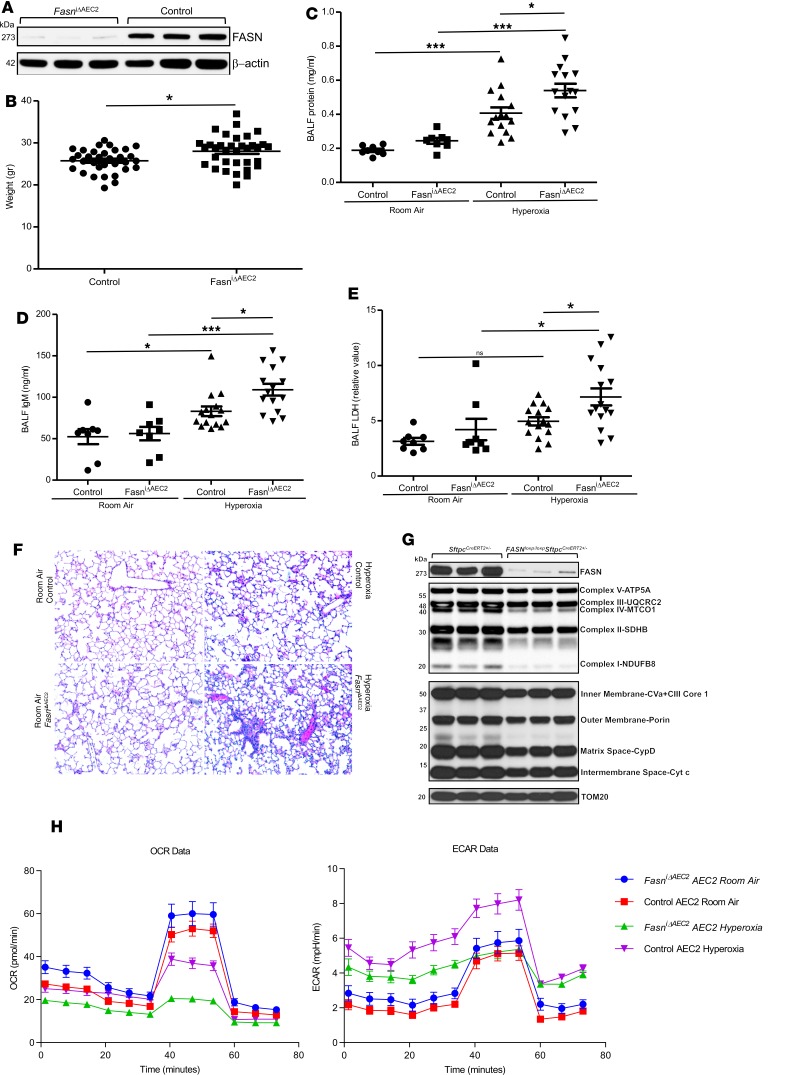
Figure 4. Mice deficient in FASN in alveolar epithelial cells are more susceptible to lung injury after hyperoxic exposure.
(A) Western blot analysis for FASN in lungs from FASNloxp/loxp SftpcCreERT2+/– (FasniΔAEC2) and SftpcCreERT2+/– (control) mice with β-actin loading control (n = 3 per group). (B) Weight (n = 33 for control and n = 34 for FasniΔAEC2 mice, Mann-Whitney U test, *P < 0.05). (C) Bronchoalveolar lavage fluid (BALF) protein levels from FasniΔAEC2 and control mice after 48 hours of exposure to >95% oxygen or room air (mg/mL, n = 8 per group for room air and n = 15 per group for hyperoxia, ANOVA with Tukey’s post hoc correction, ***P < 0.001, *P < 0.05; similar results were obtained from at least 2 independent experiments). (D) BALF IgM levels from FasniΔAEC2 and control mice after 48 hours of exposure to >95% oxygen or room air (ng/mL, n = 8 per group for room air and n = 15 per group for hyperoxia, ANOVA with Tukey’s post hoc correction, ***P < 0.001, *P < 0.05, similar results were obtained from at least 2 independent experiments). (E) BALF lactate dehydrogenase (LDH) levels after 48 hours of hyperoxia or room air (relative value, n = 8 per group for room air and n = 15 per group for hyperoxia, ANOVA with Tukey’s post hoc correction, *P < 0.05, similar results were obtained from at least 2 independent experiments). (F) Representative image of H&E-stained lungs (n = 3 per group for room air and n = 5 per group for hyperoxia; original magnification, ×20). (G) Alveolar epithelial type II (AEC2) cells were isolated from FasniΔAEC2 and control mice, and protein expression was assessed using the Total OXPHOS (ubiquinone oxidoreductase subunit B8 [NDUFB8], succinate dehydrogenase complex iron sulfur subunit B [SDHB], ubiquinol-cytochrome c reductase core protein 2 [UQCRC2], mitochondrially encoded cytochrome c oxidase 1 [MTCO1], ATP synthase subunit alpha [ATP5A]) (top) and the mitochondrial Membrane Integrity antibody cocktail (outer membrane-porin, intermembrane space-cytochrome c, inner membrane-complex VA and complex III core 1, matrix space-cyclophilin D) (bottom). TOM20 expression was used to confirm equivalent protein input (n = 3 per group). (H) Oxygen consumption rate (OCR, left) and extracellular acidification rate (ECAR, right) of isolated AEC2 cells from FASNloxp/loxp SftpcCreERT2+/– (FasniΔAEC2 AEC2) and SftpcCreERT2+/– mice (Control AEC2) exposed to room air or hyperoxia for 48 hours. All data are raw values (similar results were obtained from 2 independent experiments). Data are expressed as mean ± SEM.

