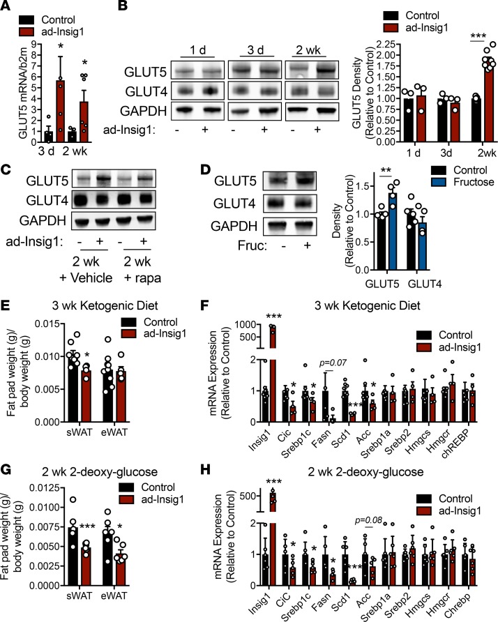
Figure 5. Chronic compensation to DNL suppression is dependent on substrate availability.
(A) GLUT5 mRNA expression in control or ad-Insig1 mice at the indicated time points on dox diet. (B) Western blot image and corresponding densitometry for GLUT4 and GLUT5 in sWAT of control or ad-Insig1 mice following the indicated duration of dox treatment in the diet (n = 3–8). (C) Representative Western blot of n = 3 from the sWAT of control or ad-Insig1 mice following 2 weeks of dox treatment with rapamycin (rapa) or vehicle injections. (D) Wild-type mice were treated with or without 20% fructose in the drinking water for 12 hours. A representative Western blot and densitometry for GLUT4 and GLUT5 in sWAT (n = 5) is shown. (E and F) Control and ad-Insig1 mice were supplied with a ketogenic diet containing dox for 3 weeks, after which (E) sWAT and eWAT weight were measured and (F) DNL gene expression was quantified (n = 5–7). (G and H) Mice were provided with 2-DG in the drinking water (0.5%) in addition to the dox diet. Following 2 weeks treatment (G) sWAT and eWAT weights were measured and (H) lipogenic enzyme mRNA was quantified (n = 5–6). All data are presented as mean ± SEM. *P < 0.05, **P < 0.01, ***P < 0.001 by 2-tailed Student’s t test. In all cases, for a given condition, representative Western blot images are from the same sample, although the target proteins may have been probed on separate membranes to facilitate clear visualization. All densitometry was conducted on samples run on the same gel.

