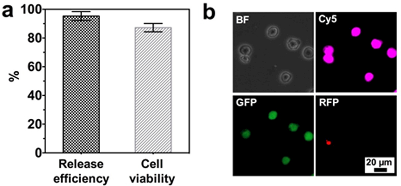
Figure 7.

(a) Release efficiency and viability of released cancer cells from NSHGMS. (b) Live (green)/Dead (red) staining of captured and released MCF7 cells. MCF7 cells were pre-stained with CellTracker™ Deep Red before spiking into blood (1000 cells/mL). Live MCF7 cells show co-localization of pink and green fluorescent signals.
