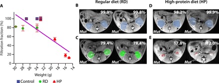
Fig. 4. In vivo FF results for MMA mice.

(A) FF calculated by taking the time-averaged FF for 10 images. The FF is defined as the percentage of kidney pixels with >20% of maximum contrast, and the SD in FF is calculated by comparing the 10 time points used. RD and HP Mut+/− controls display FF of ~100% (n = 5), whereas HP Mut−/− mice show the lowest FF, indicating that FF could be a CEST MRI metric for detecting renal disease. The blue, green, and red colors in the perfusion maps are used to denote the Mut+/−, RD Mut−/−, and HP Mut−/− mice, respectively. The decrease in FF was in accordance with a decrease in weight of the mice, which is a secondary indicator of disease progression for these mice. A linear correlation was observed between weights of mice and FF with R = 0.86 (for all groups of mice) and R = 0.9 (only considering the RD and HP Mut−/− mice; fig. S9B). (B and C) Time-averaged FF images of RD mice. (D and E) Time-averaged FF images of HP mice. RD Mut−/− mice (n = 4) have lower FF than RD Mut+/− controls but have higher FF than HP Mut−/− mice (n = 3).
