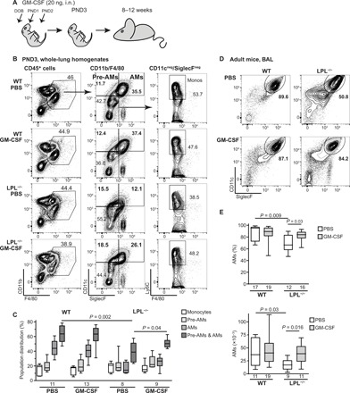
Fig. 1. Administration of rGM-CSF increases AMs in LPL−/− mice.

(A) rGM-CSF (20 ng) in 6 μl of PBS was administered via intranasal (i.n.) instillation to neonatal pups on DOB, PND1, and PND2. Mice were evaluated at indicated times after rGM-CSF administration. (B) Representative flow cytometry of whole-lung homogenates from PND3 WT and LPL−/− pups treated intranasally with PBS or rGM-CSF. (C) Quantification of the distribution of monocytes, pre-AMs, AMs, and total CD11c+ (maturing) cells in PND3 WT and LPL−/− pups. (D) Representative flow cytometry from BAL fluid obtained from adult WT and LPL−/− mice that had received neonatal rGM-CSF therapy. (E) Percentage and number of AMs recovered from BAL fluid from adult WT and LPL−/− mice that had received neonatal rGM-CSF treatment. (C and E) n of each group is provided below x axes. Data were obtained from four independent cohorts of animals. P values are determined using the Mann-Whitney U test. Kruskal-Wallis test comparing four groups revealed P = 0.0014 for AM % (top) and P = 0.015 for AM numbers (bottom). The “n” of AM numbers (bottom) in some groups is lower than AM % (top) because cell numbers in one experiment were counted manually rather than by cytometer acquisition. Only cell counts obtained by the same method (cytometer acquisition) are included in data shown here.
