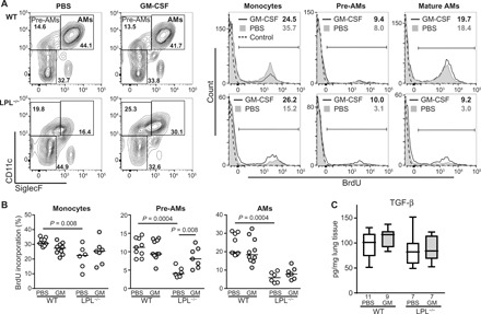
Fig. 3. rGM-CSF administration increased pre-AM proliferation in LPL−/− pups.

(A) Representative flow cytometry of BrdU incorporation into AMs and precursors (monocytes and pre-AMs, defined as shown) in whole-lung homogenates from PND3 WT and LPL−/− pups receiving rGM-CSF therapy or PBS (control). Percentage of cells in each gate incorporating BrdU listed in the top right-hand corner. (B) Quantification of the percentage of monocytes, pre-AMs, or AMs incorporating BrdU in PND3 WT or LPL−/− pups receiving neonatal rGM-CSF therapy (gray symbols) or PBS (control; open symbols). Each symbol represents one animal. Data are combined from three independent experiments. Line shows median value. P values are determined using the Mann-Whitney U test. (C) TGF-β concentration in whole-lung homogenates from PND3 WT and LPL−/− pups receiving neonatal rGM-CSF therapy (gray bars) or PBS (open bars). Line shows the median value. n of each group is listed below the x axis. Data are from two independent cohorts of animals.
