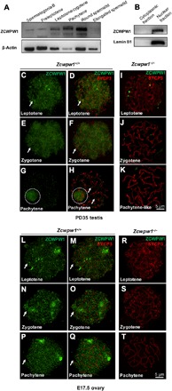
Fig. 1. ZCWPW1 expression and dynamic localization in meiotic germ cells.

(A) Western blotting of ZCWPW1 in isolated male germ cells, demonstrating that the Zcwpw1 expression level increased from the leptotene stage to the round spermatid stage and then disappeared in the elongated spermatid. β-Actin was used as the control. (B) Western blotting of ZCWPW1 in the cytoplasmic and nuclear fractions of PD35 wild-type testes shows that ZCWPW1 was only expressed in the nuclei. Lamin B1 was used as the marker of nuclear fractions. (C to K) Chromosome spreads of spermatocytes from the testes of PD35 Zcwpw1+/+ and Zcwpw1−/− males immunostained for ZCWPW1 (green) and SYCP3 (red). ZCWPW1 was diffuse (arrows) from the leptotene to zygotene stages (C to F, arrows). In pachytene cells, ZCWPW1 was localized in the XY body (G and H, white dashed circles). (I to K) In Zcwpw1−/− spermatocytes, no ZCWPW1 signal was detected. (L to T) Chromosome spreads of oocytes from E17.5 Zcwpw1+/+ and Zcwpw1−/− ovaries immunostained for ZCWPW1 (green) and SYCP3 (red). ZCWPW1 was diffuse (arrows) from the leptotene to pachytene stages (L to Q, arrows). (R to T) In Zcwpw1−/− oocytes, no ZCWPW1 signal was detected.
