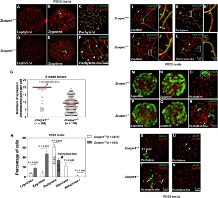
Fig. 3. Disrupted chromosomal synapsis in Zcwpw1−/− spermatocytes.

(A to F) Chromosome spreads of spermatocytes from the testes of PD35 wild-type (A to C) and Zcwpw1−/− (D to F) males were immunostained for SYCP1 (green) and SYCP3 (red). Arrows indicate synapsed chromosomes, and arrowheads indicate the single chromosome. (G). The numbers of synapsed chromosome pairs in wild-type spermatocytes and Zcwpw1−/− spermatocytes. In Zcwpw1−/− spermatocytes, the average number of synapsed chromosome pairs was 8. (H) Frequencies of meiotic stages in Zcwpw1+/+ and Zcwpw1−/− spermatocytes. The numbers marked in the bars represent the percentage of cells at indicated meiosis stage. For each genotype, three mice were analyzed. P values were calculated by Student’s t test. (I to L) SIM images of spermatocyte chromosome spreads immunostained for SYCP3 (red) and N-SYCP1 (green) from PD25 testes. Arrows indicate the synapsed region, and arrowheads indicate the AEs. (I’ to L’) Magnified views of the synapsed region show that N-SYCP1 was localized in the central region of SCs in a continuous pattern (arrows). (M to R) Chromosome spreads of spermatocytes from Zcwpw1+/+ (M to O) and Zcwpw1−/− (P and Q) males were immunostained for SYCP3 (red) and γH2AX (green). Representative images of spermatocytes at the leptotene, zygotene, and pachytene stages are shown. (S and T) Zcwpw1+/+ and Zcwpw1−/− cells immunostained for SYCP3 (red) and breast cancer 1 (BRCA1; green). Representative images of spermatocytes at pachytene (S, arrow indicating XY body) and pachytene-like (T, arrows indicating BRCA1 signal) stages are shown. (U and V) Zcwpw1+/+ and Zcwpw1−/− spermatocytes immunostained for SYCP3 (red) and MLH1 (green, arrows). Zcwpw1−/− spermatocytes lack MutL homolog 1 (MLH1) signal (V).
